
Which comes for me to find, how to make a person smarter:
Eat the right foods, and learning new things.
If babies do that, why can't we?

An adult human brain has more than 100 billion neurons. The achievement of such complexity is even more astounding when one considers that during the first few weeks after fertilization many of the sense organs are not even connected to the embryonic processing centers of the brain. During fetal brain, neurons must be generated in the right quantity and location. The axons that propagate(spread) from them must select the correct pathway to their target and finally make the right connection.

How do such precise neural links form? One idea holds that the brain wires itself as the fetus develops, in a manner analogous(similar) to the way a computer is manufactured, that is, the chips and components to the analogy, a flip of a biological switch at some point in prenatal life turns on the computer... This notion would imply that the brain's entire structure is recorded in a set of biological blueprints -- presumably DNA-- and that the organs begins to work only after the wiring is essentially complete.
Thus to believe, your brain is designed according to your DNA given from mommy and daddy. I said design, not "living from", which means after you're out of the uterus, you design your own brain...
Which brings me to ask: Could a fetal's brain structure tell a person's DNA physical appearance? Can a parent truly morph their own offspring?
This would be fascinatingly complex, and if this is indeed true, designer babies would cost a million.

The brain becomes bigger because neurons grow in size, and the numbers of axons and dendrites as well as the extent of their connections increase Human are orginally born with almost all the neurons they will ever have. The mass of the brain of a fetus is 1/4 of the adult brain.

Indeed, several observations during the past few decades have shown that babies who spent most of their first year of life lying in their cribs developed abnormally slowly. Some of these infants could not sit up at 21 months of age, and fewer than 15 percent could walk by about the age of three.
Children must be stimulated through touch, speech, and images-- to develop fully. Some people favor enriched environments for young children, yet current studies provide no clear evidence that such extra stimulation is helpful.
As a first step toward understanding the process, neurobiologists have focused on the development of the visual system in other animals, especially during the neonatal stages. It is easy under the conditions that prevail (win) at that stage to control visual experience and observe behavioral response to small changes. Furthermore, the mammalian eye differs little from species to species. Another physiological fact makes the visual system a productive object of study: its neurons are essentially the same as neurons in other parts of the brain. For these reasons, the results of such studies are very likely to be applicable to the human nervous system as well.
The visual system is the part of a central nervous system which gives organisms the ability to process visual details, as well as detect and interpret information from the eyes...

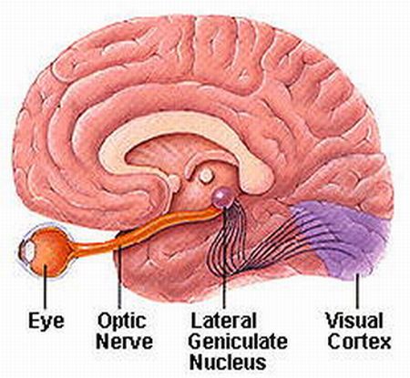
The cells of the lateral geniculate nucelus then sends the visual information to specific neurons located in what is called layer 4 of the (six layer) primary visual cortex, located at the edge of the brain's back and head. This cortical region occupies the occipital lobe in each cerebral hemisphere.


Within the lateral geniculate nucelus, retinal ganglion cell axos from each eyes are strictly segregated: the axons of one eye alternate with those from the otehr and thus form a series of eye-specific layers. The axons from the lateral geniculate nucelus terminate (end) in restricted patches within cortical layer 4. The patches corresponding to each eye interdigitate (interlock like the fingers of two clasped hands) with one another to form structures termed ocular dominance columns.
To establish such a network during development, axons must grow long distances, because the target structures form in different regions. The retinal ganglion cells are generated within the eye. Where they receive info. from the eyes and from the retinal ganglion membrane...

The diencephalon form during embryonic structure, in the same time as the shaping the lateral geniculate neurons...